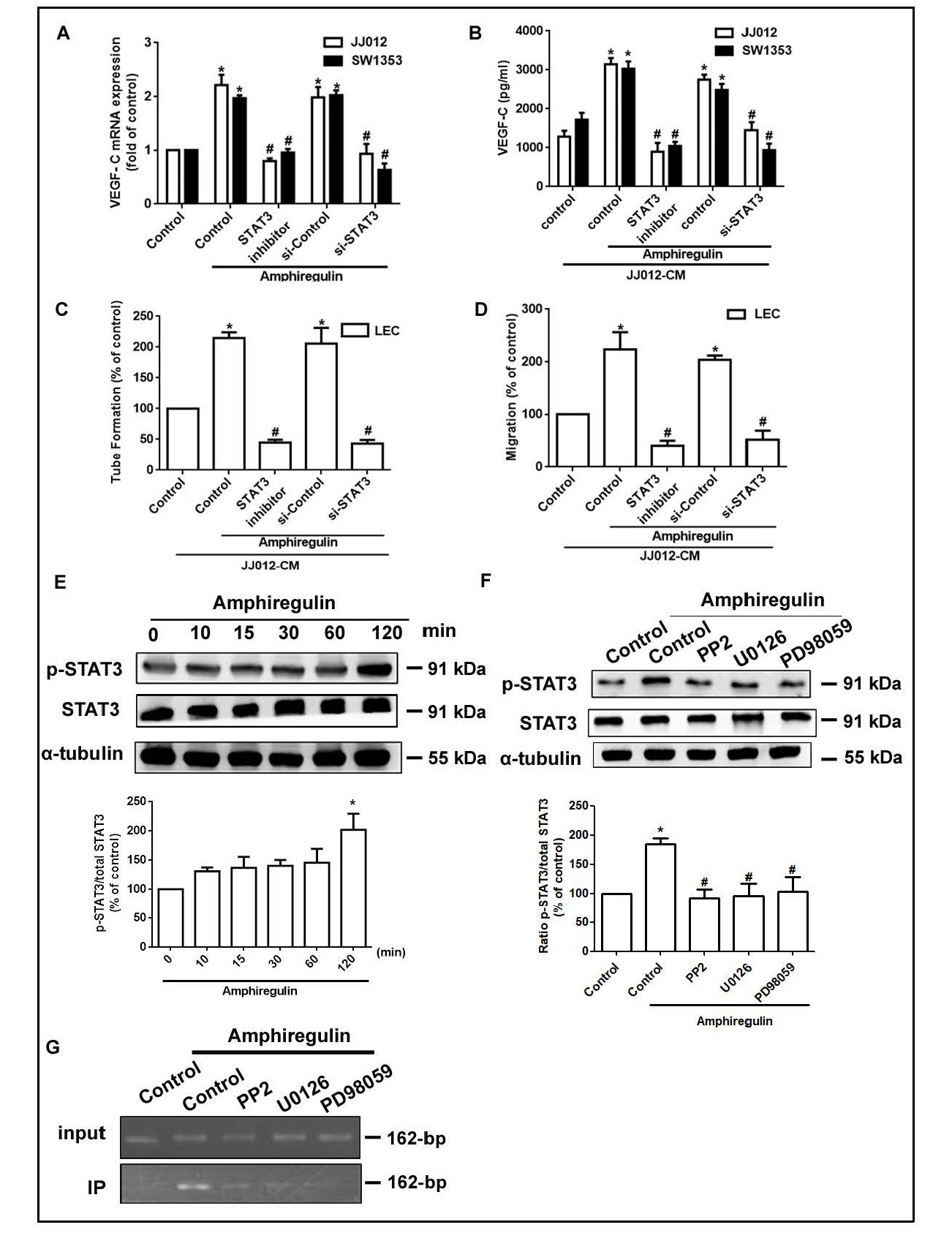
Fig. 4. AR induced VEGF-C expression and lymphangiogenesis in human chondrosarcoma cells via STAT3 activation. Clls were pretreated with a STAT3 inhibitor for 30 min or transfected with siRNAs for 24 h, then stimulated with AR (50 μg/ml) for 24 h. VEGF-C expression was examined by qPCR (A) and ELISA (B). The medium was collected as CM and then applied to the LECs. Capillary-like structure formation and LEC migration was examined by tube formation and Transwell assay, respectively (C and D). (E) JJ012 cells were incubated with AR (50 μg/ml) for 120 min, and STAT3 phosphorylation was determined by Western blotting. (F) JJ012 cells were pretreated with inhibitors as indicated, then incubated with AR for 60 min and analyzed by Western blotting with STAT3 antibodies. Western blot data were normalized to STAT3 as the loading control. (G) Cells were pretreated with PP2, U0126 and PD98059 for 30 min, then stimulated with AR (50 μg/ml), and the chromatin immunoprecipitation assay was performed. Chromatin was immunoprecipitated with anti-STAT3 antibody. One percent of the precipitated chromatin was assayed to verify equal loading (input). Results are expressed as the mean ± SEM. *P<0.05 compared with controls; #P<0.05 compared with the AR-treated group.